Review Article
Bruxism: Its multiple causes and its effects on Dental Implants: A Review

Sameera Singh Deo1, Devinder Preet Singh1* and Nitya Dogra2
1Dr. Harvansh Singh Judge Institute of Dental Sciences, Sector 25, Panjab University, Chandigarh, India
2Senior Consultant, Dr. Sabherwal’s Dental Centre, New Delhi, India
*Address for Correspondence: Dr. Devinder Preet Singh, Mohali Medical Centre, Phase 2, Opposite Bassi Cinema, Mohali, Punjab, India-160055, Tel: +919316557350; E-mail: [email protected]
Dates: Submitted: 18 April 2017; Approved: 10 May 2017; Published: 12 May 2017
How to cite this article: Sameera Singh D, Singh DP, Nitya D. Bruxism: Its multiple causes and its effects on Dental Implants: A Review. J Oral Health Craniofac Sci. 2017; 2: 057-063. DOI: 10.29328/journal.johcs.1001012
Copyright License: 2017 Sameera Singh D, et al. This is an open access article distributed under the Creative Commons Attribution License, which permits unrestricted use, distribution, and reproduction in any medium, provided the original work is properly cited.
Keywords: Dental implants; Implant-supported dental prosthesis; Mouth rehabilitation; Tooth diseases
ABSTRACT
The rehabilitation of partially or completely edentulous patients with implant supported prostheses has been widely used, achieving high success rates. However, many studies consider the presence of bruxism as a contraindication for this treatment modality. The purpose of this study was to review the literature and identify risk factors in implant supported rehabilitation planning in subjects with bruxism. The rehabilitation of bruxers using implant supported prostheses, using implants with adequate length and diameter, as well as proper positioning, seems to be a reliable treatment with reduced risks of failure. Bruxism control through the use of a night guard by rigid occlusal stabilization appliance, relieved in the region of implants, is highly indicated. Although it is clear that implant supported rehabilitation of patients with bruxism requires adequate planning and follow-up, well-designed randomized controlled trials are needed to provide reliable evidence on the long-term success of this treatment modality.
INTRODUCTION
Bruxism is a movement disorder of the masticatory system that is characterized among others by teeth grinding and clenching during sleep as well as during wakefulness [1,2]. Bruxism has prevalence in the general adult population of about 10% and is usually regarded as one of the possible causative factors for temporomandibular pain, tooth wear in the form of attrition and loss of dental implants [3]. These possible musculoskeletal and dental consequences of bruxism illustrate the clinical importance of this disorder. Most significantly, it should be kept in mind that there is still a lack of agreement about the definition of bruxism which makes it sometimes difficult to unequivocally interpret the available evidence.
The most important factor in implant longevity, i.e. clinically successful implant treatment is the formation of a direct interface between the implant and the bone, without intervening soft tissue, a process called “osseointegration”. Osseointegrated dental implants represent an advance in modern odontology which has become a great option for the rehabilitation of missing single teeth in partially or completely edentulous patients. Despite the very high success rates, complications associated with implant treatment may occur [4]. Early loading failure may affect 2% to 6% of implants, and as many as 15% of restorations failure as a result of this problem [5,6]. Excess load on a final restoration after successful implant integration can result in failure of the implant itself. Therefore, it is important to clarify the risk factors for failure of implant prostheses in order to further improve the good success rate.
The consequences of overload of dental implants can be divided into two groups: biological and biomechanical complications [7]. Biological complications can be divided into early failures and late failures [8]. In case of early failures, osseointegration was insufficient: the implant is lost before the first prosthetic loading. Late biological failures are characterized by pathological bone loss after full osseointegration was obtained at an earlier stage [9]. They are associated with overload. Some insight into bone physiology is needed for a proper understanding of these mechanisms [10].
In case of biomechanical complications, one or more components of an implant system failure are fracture of an implant itself, loosening or fracture of connecting screws or abutment screws, loosening or excessive wear of mesostructural components in overdentures and excessive wear or fracture of suprastructural porcelain or acrylic teeth [11,12]. Bragger et al. recognized a causal relation between bruxism and fracture of the suprastructure, but they could not show the relation between bruxism and failure of the implant itself.
On the other hand, Engel et al. [14], suggested that bruxism never affected the marginal bone loss of the dental implant. From these studies, it is difficult to conclude that bruxism is a risk factor for dental implants. However, in contrast, a few research data on the influence of bruxism on dental implant outcome is present, which suggest that bruxism may significantly increase both the implant failure rate and the rate of mechanical and technical complications of implant supported restorations [15,16].
In this article, the available evidence for a possible cause-and-effect relationship between bruxism and implant failure is discussed. Further, the possibility of clinical management of implant prostheses using an alteration of occlusal materials in the suprastructure and night guard in patients with bruxism is being presented.
AETIOLOGY OF BRUXISM
The literature which is so far published about the aetiology of bruxism, is often difficult to interpret. In part, this is because of the persisting disagreement about the definition and diagnosis of this disorder. However, there is consensus about the multifactorial nature of the aetiology of bruxism. Besides peripheral (viz. morphological) factors, central (viz. pathophysiological and psychosocial) factors can be distinguished. In the past, morphological factors, like occlusal discrepancies and deviations in the anatomy of the bony structures of the orofacial region have been considered the main causative factors for bruxism [17].
Nowadays, these factors are thought to play only a small role, if at all. Recent focus is more on the pathophysiological factors. For example, bruxism has been suggested to be part of a sleep arousal response, the oral motor event either preceding or following the arousal. In addition, bruxism appears to be modulated by various neurotransmitters in the central nervous system. More specifically, disturbances in the central dopaminergic system have been described in relation to bruxism. Further, factors like medication, (illicit) drugs, genetics, trauma, and neurological and psychiatric diseases may be involved in the aetiology of bruxism. Psychosocial factors like stress and personality are frequently mentioned in relation to bruxism as well. However, research to these factors comes to equivocal results and needs further attention. Taken all evidence together, bruxism seems to be mainly regulated centrally, not peripherally.
BRUXISM AS AN OCCLUSAL RISK FACTOR
In 1996, Lavigne et al. [18] proposed sleep bruxism research diagnostic criteria (SB-RDC) for polysomnographic recording, as follows: (1) a history of frequent tooth grinding occurring at least 3 nights per week for the preceding 6 months, as confirmed by a sleep partner; (2) clinical presence of tooth wear; (3) masseter muscle hypertrophy; (4) report of jaw muscle fatigue or tenderness in the morning. Bruxism is frequently considered an aetiological factor for temporomandibular disorders (TMDs), tooth wear (e.g., attrition), loss of periodontal support, and failure of dental restorations, although conflicting evidence for many of these purported aetiological relationships can be found in the literature [19].
Bruxism has also been suggested to cause excessive (occlusal) load of dental implants and their suprastructures, ultimately resulting in bone loss around the implants or even in implant failure. Therefore, bruxism is often considered a cause of concern or even a contraindication for implant treatment. In addition, many researchers use bruxism as an exclusion criterion for the selection of their participants in clinical studies concerning treatment modalities with dental implants [5].
Bruxism, other oral parafunctions, fractures of natural teeth resulting from occlusal forces, and lateral occlusal contacts on the implant prostheses were listed as important risk factors for dental implants and their suprastructures [20]. In a study of 379 patients who had used implant prostheses for many years, occlusal wear had no statistically significant impact on vertical peri-implant bone loss [21]. It was presupposed that occlusal wear was closely related to bruxism, and thus bruxism did not seem to be a risk factor for the examined variables. Tooth wear does not represent the actual/current bruxism status. It must be emphasized, of course, that bruxism is not the only cause of tooth wear and in fact is not a major factor [22].
DIAGNOSIS
The most direct way to diagnose bruxism is to evaluate the wearing of teeth (Figure 1). Nonfunctional wear facets on occlusal surfaces may occur on both natural and prosthetic teeth. Attrition of the anterior teeth appears on the incisal edge, especially in the mandible and maxillary canines, and there may be notching of the cingulum in the maxillary anterior teeth. Isolated wear of an anterior tooth is not as much of a concern if all posterior teeth contacts can be eliminated in excursions.
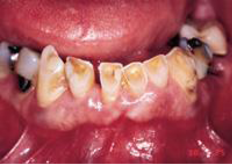
Figure 1: Tooth wear is the best method to evaluate bruxism. The “pathway of destruction” informs the dentist about parafunctional activity. The posterior wearing of teeth indicates a loss of anterior guidance.
Tooth wear is particularly significant when found in the posterior region. Posterior wear patterns are more difficult to manage because these are usually related to a loss of anterior guidance in excursive movements. If the posterior teeth are in contact during mandibular excursions, greater forces are generated [23]. Consequently, prior to restoration with an implant retained prosthesis, the occlusal plane and anterior incisal guidance may need to be restored to eliminate posterior contacts during mandibular excursions.
Patients who brux often exhibit mandibular excursions that do not correspond to border movements of the mandible. As a result, the occlusal wear is very specific and primarily on one side of the arch, or even on only a few teeth. When a habit is regularly repeated and persists after the stimuli ceases, it may be called an engram. Hence, an engram is a definite and permanent trace left by a past stimulus, [24] and usually remains after treatment. If the restoring dentist re-establishes incisal guidance on teeth severely affected by an engram bruxing pattern, the incidence of complications on these teeth will be increased [25].
The most common complications associated with teeth restored in this “pathway of destruction” are porcelain fracture, uncemented prostheses, and root fracture [26]. The amount of wear on teeth is a measure of this condition, as well as its severity (mild, moderate, or severe). Severe bruxism modifies normal masticatory forces in magnitude (higher bite forces), duration (hours rather than minutes), direction (lateral rather than vertical), type (shear rather than compression), and magnification (4 to 7 times normal) [27].
IMPLANT FATIGUE FRACTURES
The increase in force magnitude and duration on implants as a result of bruxism is a significant problem. Materials have a fatigue curve, which is related to the intensity and frequency of the force [26]. There is a force of sufficient magnitude that one cycle causes a fracture (e.g. karate blow to a piece of wood). However, if a force of lower magnitude repeatedly strikes an object, it will also fracture. The wire coat hanger that is bent does not break the first time, but repeated bending will fracture the material. This is not because the last bend was more forceful, but because of fatigue.
A bruxing patient is at greater risk for implant fracture over time because the magnitude of the force will increase as the muscles become stronger and the number of cycles accumulates. The chance of an untoward outcome will increase if the force cannot be reduced in intensity and duration. Therefore, once the dentist has identified the source(s) of additional force on the implants, the treatment plan must be altered in an attempt to minimize the adverse effect on the alveolar bone, implant, and final restoration.
How to reduce stress on implants?
Rehabilitation planning
When an implant supported rehabilitation is considered in bruxers, occlusal examination is essential. Premature and posterior contacts during mandibular excursions increase the potential for excessive loads on teeth and implants [23]. Thus, it is important to emphasize the importance of clinical evaluation and accurate anamnesis to identify the disease and hence, to improve the longevity of implant-supported rehabilitation treatment [29]. Regarding doubts about the load time determination, though a recent systematic review [30], has evaluated the load protocols for dental implants and concluded that there is no predictability in the use of immediate or early loading on dental implants, it is recommended to use caution when using any of these techniques, especially in bruxers.
Although there is no conclusive scientific evidence that bruxism causes overload on dental implants and their superstructure, professionals should proceed cautiously when planning implant supported restorations in bruxers, mainly due to the severity of possible complications. All preventive measures should be aimed at minimizing the forces that are applied to implants [5]. A bruxism patient presents an increased risk of implant fracture over time since the magnitude of forces increases as the muscles become stronger. Therefore, when a source of additional load on the implant is identified, the treatment plan must be changed to minimize adverse effects on the alveolar bone, implant and definitive restoration [31].
OCCLUSAL DEVICES
While a number of authors have reported the effectiveness of hard occlusal stabilization devices, minimizing the damage caused by parafunction on the oral tissues, clinical results have shown no differences compared to other types of devices [32]. The only exception is soft splints. Studies have indicated that soft splints can increase muscular activity in some patients with bruxism, [33] although the literature is still controversial [34]. A recent systematic review showed that rigid occlusal appliances can be effective in controlling pain in patients with temporomandibular disorders [35]. The use of soft occlusal splints led to a feeling of muscle fatigue that was accompanied by a decrease in bite force after removal of the device, while the use of rigid devices on natural teeth did not cause tiredness or modification of the bite force.
When considering only natural teeth, nightguard occlusal splints used on the upper arch can be a useful diagnostic tool in evaluating occlusal disharmony and its relationship with sleep bruxism. These devices also promote centric occlusal contacts along the arch and posterior disocclusion during the anterior guidance at all mandibular excursions and should be made of acrylic resin with a thickness of 0.5 to 1 mm on the occlusal surface [35]. If the occlusal splint becomes worn in a month, the influence of occlusion in bruxism should be directly observed in the patient. In this case, an occlusal adjustment should have little influence on bruxism reduction, as a proper occlusion provided by the splint failed to reduce the habit [36].
Number and location of implants
When rehabilitating bruxers, calculation of the number of implants is mandatory since most authors have recommended more implants than necessary to obtain favorable biomechanics [36,37]. This recommendation is justified by studies that indicate a reduction of forces received on each implant individually when the number of implants is increased [38]. Nevertheless, considering the financial costs and the irreversible nature of the placement of a greater number of implants, a careful clinical decision making process should be built into the treatment plan.
Diameter, length, and connection of implants
The use of implants with a diameter as large as possible should be considered, as well as the longest length allowed by the remaining bone to reduce stress in the cortical bone.
PROSTHESIS DESIGN
Prostheses should be designed with the purpose of improving the stress distribution on implants, i.e., implants must be installed perpendicularly to the curves of Spee and Wilson to favor direct contacts generated during the vertical function to the long axis of the implants [39]. Occlusal contacts must be precise on the antagonist arch and carefully informed to laboratory technicians in all laboratory steps [39]. The occlusal surfaces of posterior teeth can be reduced from the palatal surface of maxillary implants or the buccal surface of mandibular implants to prevent excessive lateral forces to reduce tension during chewing, leaving more space for the tongue and cheek. The cusp slope reduction of antagonistic natural teeth has often been indicated to improve vertical force distribution on implants [33].
CONCLUSION
The rehabilitation of bruxism patients through the use of implants is a feasible alternative when implants present adequate length and diameter and correct positioning, reducing the risk of treatment failure. However, control of bruxism manifestations is important, and the use of a hard occlusal stabilization splint relieved in the region of the implants during sleep is recommended. In addition, before any treatment is conducted, patients should be warned about the need for regular maintenance to avoid complications and accept the possibility of technical complications that may generate additional costs of maintenance in either implant supported or implant retained crowns in partial or complete dentures.
REFERENCES
- Thorpy MJ. International classification of sleep disorders: diagnostic and coding manual. Rochester, MN: Allen Press. 1990; 142-185.
- Okeson JP. Orofacial pain. Guidelines for assessment, diagnosis, and management. Chicago, Il: Quintessence Publishing Co. Inc. 1996.
- Lobbezoo F, Van Der Zaag J, Visscher CM, Naeije M. Oral kinesiology: A new postgraduate programme in the Netherlands. J Oral Rehabil. 2004; 31: 192-198. Ref.: https://goo.gl/PWhg2E
- Lobbezoo F, Naeije M. Bruxism is mainly regulated centrally, not peripherally. J Oral Rehabil. 2001; 28: 1085-1091. Ref.: https://goo.gl/qt7T1Q
- Lobbezoo F, Brouwers JEIG, Cune MS, Naeije M. Dental implants in patients with bruxing habits. J Oral Rehabil. 2006; 32: 152-159. Ref.: https://goo.gl/56Lp4h
- Kato T, Thie NM, Montplaisir JY, Lavigne GJ. Bruxism and orofacial movements during sleep. Dent Clin North Am. 2001; 45: 657-684. Ref.: https://goo.gl/tHNomI
- Kato T, Dal-Fabbro C, Lavigne GJ. Current knowledge on awake and sleep bruxism: overview. Alpha Omegan. 2003; 96: 24-32. Ref.: https://goo.gl/OlGWjs
- Kato T, Thie NM, Huynh N, Miyawaki S, Lavigne GJ. Topical review: sleep bruxism and the role of peripheral sensory influences. J Orofac Pain. 2003; 17: 191-213. Ref.: https://goo.gl/o1t9JK
- De Laat A, Macaluso GM. Sleep bruxism as a motor disorder. MovDisord. 2002; 17: 67-69. Ref.: https://goo.gl/AAaMNx
- Lavigne GJ, Kato T, Kolta A, Sessle BJ. Neurobiological mechanisms involved in sleep bruxism. Crit Rev Oral Biol Med. 2003; 14: 30-46. Ref.: https://goo.gl/tnhjGt
- Sari S, Sonmez H. The relationship between occlusal factors and bruxism in permanent and mixed dentition in Turkish children. J Clin Pediatr Dent. 2001; 25: 191-194. Ref.: https://goo.gl/BE46tA
- Giffin KM. Mandibular adaptive reposturing: the aetiology of a common and multifaceted autodestructive syndrome. Gen Dent. 2003; 51: 62-67. Ref.: https://goo.gl/rLUntN
- Manfredini D, Landi N, Romagnoli M, Bosco M. Psychic and occlusal factors in bruxers. Aust Dent J. 2004; 49: 84-89. Ref.: https://goo.gl/vyBFo0
- Manfredini D, Landi N, Tognini F, Montagnani G, Bosco M. Occlusal features are not a reliable predictor of bruxism. Minerva Stomatol. 2004; 53: 231-239. Ref.: https://goo.gl/OGVovS
- Chrcanovic BR, Kisch J, Albrektsson T, Wennerberg A. Bruxism and dental implant failures: a multilevel mixed effects parametric survival analysis approach. J Oral Rehabil. 2016; 43: 813-823. Ref.: https://goo.gl/ASDWbJ
- Chrcanovic BR, Kisch J, Albrektsson T, Wennerberg A. Bruxism and dental implant treatment complications: a retrospective comparative study of 98 bruxer patients and a matched group. Clin Oral Implants Res. 2016. Ref.: https://goo.gl/XQpJ88
- Demir A, Uysal T, Guray E, Basciftci FA. The relationship between bruxism and occlusal factors among seven- to 19-year-old Turkish children. Angle Orthod. 2004; 74: 672-676. Ref.: https://goo.gl/XFazdt
- Lavigne GJ, Rompre PH, Montplaisir JY. Sleep bruxism: validity of clinical research diagnostic criteria in a controlled polysomnographic study. J Dental Research. 1996; 75: 546-552. Ref.: https://goo.gl/SBDqVp
- Lavigne GJ, RomprePH, Montplaisir JY, Lobbezoo F. Motor activity in sleep bruxism with concomitant jaw muscle pain: a retrospective pilot study. Eur J Oral Sci.1997; 105: 92-95. Ref.: https://goo.gl/k6fjKy
- Manfredini D, Lobbezoo F. Relationship between bruxism and temporomandibular disorders: a systematic review of literature from 1998 to 2008. Oral Surg Oral Med Oral Pathol Oral Radiol Endod. 2010; 109: 26-50. Ref.: https://goo.gl/ahWgdb
- Johansson A, Johansson AK, Omar R, Carlsson GE. Rehabilitation of the worn dentition. J Oral Rehabil. 2008; 35: 548-566. Ref.: https://goo.gl/7ZEFXN
- Lobbezoo F, van der Zaag J, Naeije M. Bruxism: its multiplecauses and its effects on dental implants - an updated review. J Oral Rehabil. 2006; 33: 293-300. Ref.: https://goo.gl/MVKL6T
- Williamson EH, Lundquist DO. Anterior guidance: its effect on electromyographic activity of temporal and masseter muscles. J Prosthet Dent. 1983; 49: 816-823. Ref.: https://goo.gl/p58mhD
- Dorland’s Illustrated Medical Dictionary. WB Saunders Co: Philadelphia, Pa; 1988.
- Kois J. Manual on Functional Occlusion. Self-published: Seattle, Wash; 2002.
- Glaros AG, Rao SM. Effects of bruxism, a review of the literature. J Prosthet Dent. 1977; 38: 149-157. Ref.: https://goo.gl/Q4CKZR
- Misch CE. Dental evaluation factors of stress. In: CE Misch, ed. Contemporary Implant Dentistry. 2nd ed. St Louis, Mo: CV Mosby; 1999.
- Bidez MW, Misch CE. Clinical biomechanics in implant dentistry. In: Misch CE, ed. Contemporary Implant Dentistry. St Louis, Mo: CV Mosby; 1999: 303-316.
- Van der Zaag J, Lobbezoo F, Van der Avoort PG, Wicks DJ, Hamburger HL et al. Effects of pergolide on severe sleep bruxism in a patient experiencing oral implant failure. J Oral Rehabil.2007; 34: 317-322. Ref.: https://goo.gl/QCXiZp
- Esposito M, Grusovin MG, Achille H, Coulthard P, Worthington HV. Interventions for replacing missing teeth: different times for loading dental implants. Cochrane Database Syst Rev. 2009; 21. Ref.: https://goo.gl/vgj6Ix
- Misch CE. The effect of bruxism on treatment planning for dental implants. Dent Today. 2002; 21: 76-81. Ref.: https://goo.gl/70v08u
- Tu¨rp JC, Komine F, Hugger A. Efficacy of stabilization splints for the management of patients with masticatory muscle pain: a qualitative systematic review. Clin Oral Investig .2004; 8: 179-195. Ref.: https://goo.gl/GUJd7Q
- Okeson JP. The effects of hard and soft occlusal splints on nocturnal bruxism. J Am Dent Assoc. 1987; 114: 788-791. Ref.: https://goo.gl/7Yv1RC
- List T, Axelsson S. Management of TMD: evidence from systematic reviews and meta-analyses. J Oral Rehabil. 2010; 37: 430-451. Ref.: https://goo.gl/B6OkoA
- Thie NM, Kato T, Bader G, Montplaisir JY, Lavigne GJ. The significance of saliva during sleep and the relevance of oromotor movements. Sleep Med Rev. 2002; 6: 213-227. Ref.: https://goo.gl/KNaesf
- Narita N, Funato M, Ishii T, Kamiya K, Matsumoto T. Effects of jaw clenching while wearing an occlusal splint on awareness of tiredness, biteforce and EEG power spectrum. J Prosthodont Res. 2009; 53: 120-125. Ref.: https://goo.gl/qEkV0I
- Sarmento HR, Dantas RV, Pereira-Cenci T, Faot F. Elements of Implant-Supported Rehabilitation Planning in Patients with Bruxism. J Craniofac Surg. 2012; 23: 1905-1909. Ref.: https://goo.gl/YbcqGa
- Lindquist LW, Rockler B, Carlsson GE. Bone resorption around fixtures in edentulous patients treated with mandibular fixed tissue-integrated prostheses. J Prosthet Dent.1988; 59: 59-63. Ref.: https://goo.gl/qnTEIM
- Misch C. Dental evaluation factors of stress. In: Contemporary Implant Dentistry 2nd ed. St Louis: Mosby, 1999.