More Info
Author's satisfaction with
- Friendly and hassle-free publication process
- Less production time of articles
- Constructive peer-review
- Enhancing journal reputation
- Regular feedback system
- Quick response to authors' queries
Recently Viewed
Most Viewed
Clinical Images
Abstract
Case Report
Bone Compactness and Bad Split in the SSO (Sagittal Split Osteotomy) of the Mandible
Carmine Taglialatela Scafati* and Giuseppe Di Costanzo
Published: 14 October, 2025 | Volume 10 - Issue 1 | Pages: 001-006
Sagittal split osteotomy (SSO) of the mandible is still an important part of orthognathic surgery, but “bad split” fractures that happen during the surgery are still a big problem. This report describes a rare case of bilateral bad split associated with highly compact mandibular bone (D1–D2 type) in a 53-year-old male undergoing mandibular advancement. The bone was too hard for the piezoelectric and rotary tools to work, and there was very little bleeding. This caused fractures in the cortex and lingual. Postoperative computed tomography confirmed the presence of highly dense bone, correlated with delayed healing and recurrence. Consistent pre-surgical CT scanning for an assessment of the density of the mandibular bone can provide essential information about the potential risk of the osteotomy procedure in the patient. Performing a careful bone quality and density examination pre-operatively will allow the surgeons to select the most appropriate instruments and surgical techniques that will fit the patient’s individual anatomy. Such a point escalates to being very significant when dealing with the geriatric population, as bone density increase can not only alter the fracture behavior but also the recovery process during and after surgery. The inclusion of regular CT-based bone density evaluations into the preoperative routine not only facilitates the anticipation of surgical difficulties but also results in safer and more efficient osteotomies.
Read Full Article HTML DOI: 10.29328/journal.johcs.1001051 Cite this Article Read Full Article PDF
Keywords:
Sagittal split osteotomy; Bad split; Mandibular bone density; CT evaluation; D1 bone; Orthognathic surgery
References
- Guernsey LH, DeChamplain RW. Sequelae and complications of the intraoral sagittal osteotomy in the mandibular rami. Oral Surg Oral Med Oral Pathol. 1971;32(2):176-92. Available from: https://doi.org/10.1016/0030-4220(71)90221-0
- Van Merkesteyn JP, Groot RH, van Leeuwaarden R, Kroon FH. Intra-operative complications in sagittal and vertical ramus osteotomies. Int J Oral Maxillofac Surg. 1987;16(6):665-70. Available from: https://doi.org/10.1016/s0901-5027(87)80050-4
- Kriwalsky MS, Maurer P, Veras RB, Eckert AW, Schubert J. Risk factors for a bad split during sagittal split osteotomy. Br J Oral Maxillofac Surg. 2008;46(3):177-9. Available from: https://doi.org/10.1016/j.bjoms.2007.09.011
- MacIntosh RB. Experience with the sagittal osteotomy of the mandibular ramusa 13-year review. J Maxillofac Surg. 1981;9(3):151-65. Available from: https://doi.org/10.1016/s0301-0503(81)80036-7
- Van Merkesteyn JP, Groot RH, van Leeuwaarden R, Kroon FH. Intra-operative complications in sagittal and vertical ramus osteotomies. Int J Oral Maxillofac Surg. 1987;16(6):665-70. Available from: https://doi.org/10.1016/s0901-5027(87)80050-4
- Mensink G, Verweij JP, Frank MD, Bergsma JE, van Merkesteyn JP. Bad split during bilateral sagittal split osteotomy of the mandible with separators: a retrospective study of 427 patients. Br J Oral Maxillofac Surg. 2013;51(6):525-9. Available from: https://doi.org/10.1016/j.bjoms.2012.10.009
- Acebal-Bianco F, Vuylsteke PL, Mommaerts MY, De Clercq CA. Perioperative complications in corrective facial orthopedic surgery: a 5-year retrospective study. J Oral Maxillofac Surg. 2000;58(7):775-80. Available from: https://doi.org/10.1053/joms.2000.7874
- Chrcanovic BR, Freire-Maia B. Risk factors and prevention of bad splits during sagittal split osteotomy. Oral Maxillofac Surg. 2012;16(2):119-27. Available from: https://doi.org/10.1007/s10006-011-0287-4
- Mehra P, Castro V, Freitas RZ, Wolford LM. Complications of the mandibular sagittal split ramus osteotomy associated with the presence or absence of third molars. J Oral Maxillofac Surg. 2001;59(8):885-8. Available from: https://doi.org/10.1053/joms.2001.25013
- Salman S, Young M, Van Sickels JE. Osteomyelitis after bilateral sagittal split osteotomy: case report and a review of the management. Oral Surg Oral Med Oral Pathol Oral Radiol Endod. 2011;111(4):442-8. Available from: https://doi.org/10.1016/j.tripleo.2010.06.003
- Teltzrow T, Kramer FJ, Schulze A, Baethge C, Brachvogel P. Perioperative complications following sagittal split osteotomy of the mandible. J Craniomaxillofac Surg. 2005;33(5):307-13. Available from: https://doi.org/10.1016/j.jcms.2005.04.005
- Guernsey LH, DeChamplain RW. Sequelae and complications of the intraoral sagittal osteotomy in the mandibular rami. Oral Surg Oral Med Oral Pathol. 1971;32(2):176-92. Available from: https://doi.org/10.1016/0030-4220(71)90221-0
- O’Ryan F. Complications of orthognathic surgery. Oral Maxillofac Surg Clin North Am. 1990;2(1):93-601.
- Turvey TA. Intraoperative complications of sagittal osteotomy of the mandibular ramus: incidence and management. J Oral Maxillofac Surg. 1985;43(7):750-4. Available from: https://doi.org/10.1016/s0278-2391(85)80028-8
- Veras RB, Kriwalsky MS, Hoffmann S, Maurer P, Schubert J. Functional and radiographic long-term results after bad split in orthognathic surgery. Int J Oral Maxillofac Surg. 2008;37(7):760-6. Available from: https://doi.org/10.1016/j.ijom.2008.04.010
- Lee SY, Yang HJ, Han JJ, Hwang SJ. Effect of perioperative buccal fracture of the proximal segment on postoperative stability after sagittal split ramus osteotomy. J Korean Assoc Oral Maxillofac Surg. 2013;39(5):217-23. Available from: https://doi.org/10.5125/jkaoms.2013.39.5.217
- Epker BN, Fish LC. Dentofacial deformities. St. Louis: Mosby; 1986;1:232-4. Available from: https://search.library.ucdavis.edu/discovery/fulldisplay?vid=01UCD_INST:UCD&tab=Everything&docid=alma990006424270403126
- Gilles R, Couvreur T, Dammous S. Ultrasonic orthognathic surgery enhancements to established osteotomies. Int J Oral Maxillofac Surg. 2013;42(8):981-7. Available from: https://doi.org/10.1016/j.ijom.2012.12.004
- Akhtar S, Tuinzing DB. Unfavorable splits in sagittal split osteotomy. Oral Surg Oral Med Oral Pathol Oral Radiol Endod. 1999;87(3):326-7. Available from: https://doi.org/10.1016/s1079-2104(99)70230-1
- Hounsfield GN. Computerized transverse axial scanning tomography. 1. Description of system. Br J Radiol. 1973;46(552):1016-22. Available from: https://doi.org/10.1259/0007-1285-46-552-1016
- Misch CE. Second vital implant criterion. Dental Today. 1988 :39-40.
- Trauner R, Obwegeser H. Zur Operationstechnik bei der Progenie und anderen Unterkieferanomalien. Dtsch Zahn Mund Kieferheilkd. 1955;23:11-26. Available from: https://www.scirp.org/reference/referencespapers?referenceid=1257402
- Dal Pont G. Retromolar osteotomy for the correction of prognathism. J Oral Surg Anesth Hosp Dent Serv. 1961;19:42-7. Available from: https://pubmed.ncbi.nlm.nih.gov/13719390/
- Shapurian T, Damoulis PD, Reiser GM, Griffin TJ, Rand WM. Quantitative evaluation of bone density using the Hounsfield index. Int J Oral Maxillofac Implants. 2006;21 https://pubmed.ncbi.nlm.nih.gov/16634501/
- Kingsmill VJ, Boyde A. Mineralisation density of human mandibular bone: quantitative backscattered electron image analysis. J Anat. 1998;192(Pt 2):245-56. Available from: https://doi.org/10.1046/j.1469-7580.1998.19220245.x
- Simpson W. Problems encountered in the sagittal split operation. Int J Oral Surg. 1981;10:81-6. Available from: https://doi.org/10.1016/s0300-9785(81)80016-6
- Muto T, Akizuki K, Tsuchida N, Sato Y. Modified intraoral inverted L osteotomy: a technique for good visibility, greater bony overlap, and rigid fixation. J Oral Maxillofac Surg. 2008;66(8):1309. Available from: https://doi.org/10.1016/j.joms.2007.03.034
- Landes CA, Stübinger S, Ballon A, Sader R. Piezoosteotomy in orthognathic surgery versus conventional saw and chisel osteotomy. Oral Maxillofac Surg. 2008;12(3):139-47. Available from: https://doi.org/10.1007/s10006-008-0123-7
- Nagadia R, Tay AB, Chan LL, Chan ES. The spatial location of the mandibular canal in Chinese: a CT study. Int J Oral Maxillofac Surg. 2011;40(12):1401-5. Available from: https://doi.org/10.1016/j.ijom.2011.07.904
- Agbaje JO, Sun Y, De Munter S, Schepers S, Vrielinck L, Lambrichts I, Politis C. CBCT-based predictability of attachment of the neurovascular bundle to the proximal segment of the mandible during sagittal split osteotomy. Int J Oral Maxillofac Surg. 2013;42(3):308-15. Available from: https://doi.org/10.1016/j.ijom.2012.07.012
- Plooij JM, Naphausen MT, Maal TJ, Xi T, Rangel FA, Swennen G, de Koning M, Borstlap WA, Berg SJ. 3D evaluation of the lingual fracture line after a bilateral sagittal split osteotomy of the mandible. Int J Oral Maxillofac Surg. 2009;38(12):1244-9. Available from: https://doi.org/10.1016/j.ijom.2009.07.013
- Song JM, Kim YD. Three-dimensional evaluation of lingual split line after bilateral sagittal split osteotomy in asymmetric prognathism. J Korean Assoc Oral Maxillofac Surg. 2014;40(1):11-6. Available from: https://doi.org/10.5125/jkaoms.2014.40.1.11
- De Oliveira RC, Leles CR, Normanha LM, Lindh C, Ribeiro-Rotta RF. Assessments of trabecular bone density at implant sites on CT images. Oral Surg Oral Med Oral Pathol Oral Radiol Endod. 2008;105(2):231-8. Available from: https://doi.org/10.1016/j.tripleo.2007.08.007
- Song YD, Jun SH, Kwon JJ. Correlation between bone quality evaluated by cone-beam computerized tomography and implant primary stability. Int J Oral Maxillofac Implants. 2009 ;24(1):59-64. Available from: https://pubmed.ncbi.nlm.nih.gov/19344026/
- Proffit WR, White RP, Sarver DM. Contemporary Treatment of Dentofacial Deformity. St. Louis: Mosby; 2003. Available from: https://books.google.co.in/books/about/Contemporary_Treatment_of_Dentofacial_De.html?id=fyNqAAAAMAAJ&redir_esc=y
Figures:
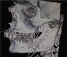
Figure 1
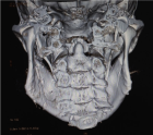
Figure 2
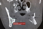
Figure 3
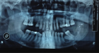
Figure 4
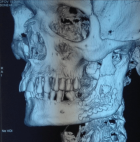
Figure 5
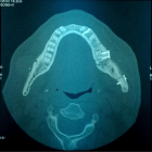
Figure 6
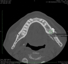
Figure 7
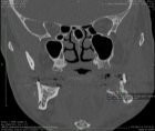
Figure 8
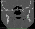
Figure 9
Similar Articles
-
Correlation between mandibular condylar position and clinical dysfunction index after bilateral sagittal split osteotomiesKalach Mussali Alberto J*,Gonzalez-Magana Fernando,Hidalgo Hector O. Correlation between mandibular condylar position and clinical dysfunction index after bilateral sagittal split osteotomies. . 2019 doi: 10.29328/journal.johcs.1001026; 4: 001-007
-
Role of orthodontist in cleft lip and palatePrashant Sharma*,Amit Kumar Khera,Pradeep Raghav. Role of orthodontist in cleft lip and palate. . 2021 doi: 10.29328/journal.johcs.1001035; 6: 008-015
-
Bone Compactness and Bad Split in the SSO (Sagittal Split Osteotomy) of the MandibleCarmine Taglialatela Scafati*,Giuseppe Di Costanzo. Bone Compactness and Bad Split in the SSO (Sagittal Split Osteotomy) of the Mandible. . 2025 doi: 10.29328/journal.johcs.1001051; 10: 001-006
Recently Viewed
-
Assessment of the Quality of Life in Patients Undergoing Radical Cystectomy with Urinary DiversionMohamed Ali Mikou,Ilyas Soufiani,Mohamed Bakouch,Hamza El-Abidi,Idriss Ziani,Imad Boualaoui,Ahmed Ibrahimi,Yassine Nouini. Assessment of the Quality of Life in Patients Undergoing Radical Cystectomy with Urinary Diversion. J Clin Med Exp Images. 2026: doi: 10.29328/journal.jcmei.1001048; 10: 44-49
-
Theranostics: A Unique Concept to Nuclear MedicineLucio Mango*. Theranostics: A Unique Concept to Nuclear Medicine. Arch Cancer Sci Ther. 2017: doi: 10.29328/journal.acst.1001001; 1: 001-004
-
Exploring the Potential of Medicinal Plants in Bone Marrow Regeneration and Hematopoietic Stem Cell TherapyUgwu Okechukwu Paul-Chima*,Alum Esther Ugo. Exploring the Potential of Medicinal Plants in Bone Marrow Regeneration and Hematopoietic Stem Cell Therapy. Int J Bone Marrow Res. 2025: doi: 10.29328/journal.ijbmr.1001019; 8: 001-005
-
Examining the Effects of High Poverty and Unemployment on Rural Urban Migration in Nigeria and its Consequences on Urban Resources and Rural DeclineTochukwu S Ezeudu*, Bilyaminu Tukur. Examining the Effects of High Poverty and Unemployment on Rural Urban Migration in Nigeria and its Consequences on Urban Resources and Rural Decline. J Child Adult Vaccines Immunol. 2024: doi: 10.29328/journal.jcavi.1001012; 8: 001-013
-
Screening for Depressive Symptoms in Clinical and Nonclinical Youth: The Psychometric Properties of the Dutch Children’s Depression Inventory-2 (CDI-2)Denise HM Bodden*,Yvonne Stikkelbroek,Daan Creemers,Sanne PA Rasing,Elien De Caluwe,Caroline Braet. Screening for Depressive Symptoms in Clinical and Nonclinical Youth: The Psychometric Properties of the Dutch Children’s Depression Inventory-2 (CDI-2). Insights Depress Anxiety. 2025: doi: 10.29328/journal.ida.1001047; 9: 028-039
Most Viewed
-
Physical Performance in the Overweight/Obesity Children Evaluation and RehabilitationCristina Popescu, Mircea-Sebastian Șerbănescu, Gigi Calin*, Magdalena Rodica Trăistaru. Physical Performance in the Overweight/Obesity Children Evaluation and Rehabilitation. Ann Clin Endocrinol Metabol. 2024 doi: 10.29328/journal.acem.1001030; 8: 004-012
-
Hypercalcaemic Crisis Associated with Hyperthyroidism: A Rare and Challenging PresentationKarthik Baburaj*, Priya Thottiyil Nair, Abeed Hussain, Vimal MV. Hypercalcaemic Crisis Associated with Hyperthyroidism: A Rare and Challenging Presentation. Ann Clin Endocrinol Metabol. 2024 doi: 10.29328/journal.acem.1001029; 8: 001-003
-
Effects of dietary supplementation on progression to type 2 diabetes in subjects with prediabetes: a single center randomized double-blind placebo-controlled trialSathit Niramitmahapanya*,Preeyapat Chattieng,Tiersidh Nasomphan,Korbtham Sathirakul. Effects of dietary supplementation on progression to type 2 diabetes in subjects with prediabetes: a single center randomized double-blind placebo-controlled trial. Ann Clin Endocrinol Metabol. 2023 doi: 10.29328/journal.acem.1001026; 7: 00-007
-
Exceptional cancer responders: A zone-to-goDaniel Gandia,Cecilia Suárez*. Exceptional cancer responders: A zone-to-go. Arch Cancer Sci Ther. 2023 doi: 10.29328/journal.acst.1001033; 7: 001-002
-
Ectopic adrenal tissue at the spermatic cordAbdallah Chaachou,Nizar Cherni,Wael Ferjaoui*,Mohamed Dridi,Samir Ghozzi. Ectopic adrenal tissue at the spermatic cord. J Clin Med Exp Images. 2022 doi: 10.29328/journal.jcmei.1001024; 6: 001-002
Contact
Select by Volume & Issue
Most Viewed Keywords
- Packed cell volume
- Prevention
- Hemoglobinuria
- Tumours of the uterine corpus
- Novel mutation
- Orthopoxvirus
- Upper molar distalization
- Periodontitis
- Bisphosphonate-Related Osteonecrosis of the Jaw (BRONJ)
- Lipoprotein apheresis
- Stevia
- Aggression
- Florfenicol
- Hemoglobinuria
- Albino mice
- Urinary microbiome
- Ischemia
- Arabian
- Antifungal activity
- Pulmonary
University/Institution
Select and search by University/Institution.
Articles by Country
Select and search by country to get related articles.


Testmonials

I would like to thank JPRA for taking this decision. I understand the effort it represents for you. I'm truly happy to have the paper published in JPRA. And I'll certainly consider JPRA for my next publications as I was satisfied of the service provided, the efficiency and promptness of the interactions we had.
Emmanuel BUSATO
Publishing with the International Journal of Clinical and Experimental Ophthalmology was a rewarding experience as review process was thorough and brisk. Their visibility online is second to none as their published articles appear in all search engines. I will encourage researchers to publish with them.
Elizabeth Awoyesuku
“The choice to submit the forensic case study to the Journal of Addiction Therapy and Research was dictated by the match between the content and the potential readership. The publication process proved to be expedient and we were provided with constructive feedback from reviewers. The final article layout is attractive and conforms to standards. All-in-all, it has been a rewarding process.”
Elisabeth H Wiig
Archives of Vascular Medicine is one of the top class journal for vascular medicine with highly interesting topics. You did a professional and great Job!
Elias Noory
Thank you very much. I think the review process and all of what concerns the administration of the publication concerning our paper has been excellent. The nice and quick answers have been very good I think.
Doris Nilsson
Journal of Pulmonary and Respiratory Research is good journal for respiratory research purposes. It takes 2-3 weeks maximum for review of the manuscript to get published and any corrections to be made in the manuscript. It needs good articles and studies to get publish in the respiratory medicine. I am really glad that this journal editors helped me to get my case report published.
Divya Khanduja
Thanks you and your colleague for the great help for our publication. You always provide prompt responses and high quality of service. I am so happy to have you working with me. Thanks again!
Diana (Ding) Dai
Service and process were excellent as was the “look” of the article when published.
Deane Waldman
Great, thank you! It was very efficient working w/ your group. Very thorough reviews (i.e., plagiarism, peer, etc.). Would certainly recommend that future authors consider working w/ your group.
David W Brett
Your services are very good
Chukwuka Ireju Onyinye
I very much appreciate the humanitarian services provided in my stead by this journal/publisher. It exhibits total absence of editorial impertinence. As an Author, I have been guided to have a fruitful experience. The editorial care is highly commendable.
Chrysanthus Chukwuma
"An amazing experience with the Journal of Advanced Pediatrics and Child Health. Very fast blind review with pertinent corrections and suggestions. I highly recommand both the journal and the editor."
Chaimae Khairoun
The submission is very easy and the time from submission to response from the reviewers is short. Correspondence with the journal is nice and rapid.
Catrin Henriksson
The Clinical Journal of Obstetrics and Gynecology is an open access journal focused on scientific knowledge publication with emphasis laid on the fields of Gynecology and Obstetrics. Their services toward us have been encouraging through their kindness and respect. Great consideration has been given to us as young budding researchers and we are very grateful for this.
Carole Assontsa
During the process your positive communication, prompt feedback and professional approach is very highly appreciated. We would like to thank you very much for your support.
Can Vuran
I do appreciate for your service including submission, analysis, review, editorial and publishing process. I believe these esteemed journal enlighten the science with its high-quality personel.
Bora Uysal
I am very much pleased with the fast track publication by your reputed journal's editorial team. It is really helpful for researchers like me from developing nations. I strongly recommend your journal for publication.
Badri Kumar Gupta
It has been a fabulous journey writing articles for your journal because of the encouragement you people provide for writers from developing nations like India. Kindly continue the same. Looking forward for a long term association.
Badareesh Lakshminarayana
Many thanks for publishing my article in your great journal and the friendly and hassle-free publication process, the constructive peer-review, the regular feedback system, and the Quick response to any queries.
Azab Elsayed Azab
I would like to thank this journal for publishing my Research Article. Something I really appreciate about this journal is, they did not take much time from the day of Submission to the publishing date. Looking forward to have more publications in future.
Ayush Chandra
Submission of paper was smooth, the review process was fast. I had excellent communication and on time response from the editor.
Ayokunle Dada
Your service is very good and fast reply, also your service understand our situation and support us to publication our articles.
Ayman M Abu Mustafa
Really good service with prompt response. Looking forward to having long lasting relationship with your journal
Avishek Bagchi
Your service is excellent. Processing and editing were very fast. I hope to publish more of my works in your journal.
Ausraful Islam
I wanna to thank Clinical Journal of Nursing Care and Practice for its effort to review and publish my manuscript. This is reputable journal. Thank you!
Atsedemariam Andualem
“It was a delightful experience publishing my manuscript with the Clinical Journal of Obstetrics and Gynecology. They offered me lots of opportunities I never had from most publishing houses and their prompt services are greatly appreciated.”
Asafo Jones
I hope to ability to make some new investigation and publish in Your Company in future.
Artur Stopyra
I like the quality of the print & overall service. The paper looks quite impressive. Hope this will attract interested readers. All of you have our best wishes for continued success.
Arshad Khan
Your big support from researchers around the world is the best appreciation from your scientific teams. We believe that there should be no barrier in science and you make it real and this motto come true.
Arefhosseinir Rafi
Your journal co-operation is very appreciable and motivational. I am really thankful to your journal and team members for the motivation and collaboration to publish my work.
Assistant Professor, UCLAS Uttaranchal University, Dehradun, India
Archna Dhasmana
I am glad to submit the article to Heighten Science Publications as it has a very smooth and fast peer-review process, which enables the researchers to communicate their work on time.
Anupam M
This is to specify that I have had an extensive and detailed interaction with the Editorial team of Annals of Clinical Gastroenterology and Hepatology, USA, lasting over a significant period of time. My interaction has been extremely pleasant, especially with Ms Allie Smith, as I find the communication quite inspiring and crystal clear. The attitude of aforesaid individuals is quite helpful and guiding in pertinent instances. It has been a commemorative journey so far working with the Journal and I hope that the symbiosis will continue, evolve and flourish in the forthcoming years. I wish the journal, related personnel and aforementioned individuals a fruitful, successful run.
New Delhi, India
Anubha Bajaj
We appreciate the fact that you decided to give us full waiver for the applicable charges and approve the final version. You did an excellent job preparing the PDF version. Of course we will consider your magazine for our future submissions and we will pay the applicable fees then.
Anna Dionysopoulou
''Co-operation of Archives of Surgery and Clinical Research journal is appreciable. I'm impressed at the promptness of the publishing staff and the professionalism displayed. Thank you very much for your support, help and encouragement.''
Anıl Gokce
Congratulations for the excellence of your journal and high quality of its publications.
Angel MARTIN CASTELLANOS
The service from the journal staff has been excellent.
Andy Smith
I was very pleased with the quick editorial process. We are sure that our paper will have great visibility, among other things due to its open access. We believe in science accessible to all.
Anderson Fernando de Souza
It was a great experience publishing through JCICM. The article has reached out to several institutions. Appreciate your professional work. Hope to work with you again
Anas Wardeh
Publishing an article is a long process, but working with your publication department made things go smoothly, even though the process took exactly 5 months from the time of submitting the article till the time I have favourable response, the missing part is the peer review details, which is essential in self auditing and future improvement, overall experience was excellent giving your understanding of the situation of lack of financial institution support.
Anas Diab
I think that Heighpubs very good. You are very helpful. Thank you for everything.
Ana Ribeiro
Regarding to be services, we note that are work with high standards of professionalism translated into quick response, efficiency which makes communication accessible. Furthermore, I believe to be much inviting for the submission of future works for publication purposes.
Amélia João Alice Nkutxi
I would like to mention that I had a wonderful experience working with HSPI. The whole process right from manuscript submission to peer review till the publication of the article was very prompt & efficient. I wish you good luck for the future.
Amarjeet Gambhir
Once I submitted the manuscript, the response time of the reviewers was very fast. The fine-tuning of the galley proof was likewise prompt. I believe the journal provide a valuable outlet to disseminate physical rehabilitation scientific knowledge to the clinical community. Respectfully. Dr. Alon
Alon
We really appreciate and thanks the full waiver you provide for our article. We happy to publish our paper in your journal. Thank you very much for your good support and services.
Ali Abusafia
It was a real pleasure working with your team. The review was done fast, and it was very clear, the editing was flawless, the article was published quickly compared to other journals, and everyone was understanding and helpful. I will gladly recommend this journal to my acquaintances in academia.
Alexandra Cozma
To the editorial team at HSPI and the Journal of Clinical Nephrology: Thank you so much for your hard work and collaboration in bringing our article to life. Your staff was responsive, flexible, and communicative and made the process smooth and easy. Thank you!
Alejandro Munoz
Dear colleagues! I am satisfied with our cooperation with you. Your service is at a high level. I hope for a future relationship. Let me know if I can get a paper version of the magazine with my articles from you. I see them on the Internet.
Aksenov V.V
"This is my first time publishing with the journal/publisher. I am impressed at the promptness of the publishing staff and the professionalism displayed. Thank you for encouraging young researchers like me!"
Ajite Kayode
I want to thank you for our collaboration. You were fast and effective with a positive spirit of teamwork. I am truly excited from our collaboration. You were like always fast, efficient and accurate. I hope that in the near future we will collaborate again.
Aikaterini Solomou
In my opinion, you provide a very fast and practical service.
Ahmet Eroglu
Great, We are too comfortable with the process including the peer review process and quality. But, the journal should be indexed in different databases such scopus.
Afework Edmealem
We really appreciate your efforts towards our article, the professional way you handle our request for exemption from charges. It was a great honor for us to publish in your magazine.
Achraf elbakkaly
I really liked the ease of submitting my manuscript in the HSPI journal. Further, the peer review was timely completed and I was communicated the final decision on my manuscript within 10 days of submission which is really appreciable. I strongly recommend all the scientists and researchers to submit their work in this journal”
Abu Bashar
My candid opinion is that the service you render is second to none. My favourite part is the prompt response to issue, really i value that.
Abiodun Akanbi Adeogun
Thank you very much for accepting our manuscript in your journal “International Journal of Clinical Virology”. We are very thankful to the esteemed team for timely response and quick review process. The editorial team of International Journal of Clinical Virology is too cooperative and well-mannered during the publication process. We are hopeful to publish many quality papers in your journal and I suggest the International Journal of Clinical Virology to all of my colleagues, researchers and friends to publish their research here.
Abdul Baset
I, Muhammad Sarwar Khan, am serving as Editor on Archives of Biotechnology and Biomedicine (ABB). I submitted an editorial titled, 'Edible vaccines to combat Infectious Bursal Disease of poultry' for publication in ABB. After submitting the manuscript; the services rendered by the management and technical personnel to handle and process the manuscript were marvelous. Plagiarism report was shared with me with complements before reviewers' comments, All steps including article processing and service charges were well taken care of keeping in view the author's interest/preference. All together, it was an encouraging and wonderful experience working with ABB personnel.
University of Agriculture, Pakistan
Muhammad Sarwar Khan
Your journal has accomplished its intended mission of providing very effective and efficient goals in dealing with submissions, conducting the reviewing process and in publishing accepted manuscripts in a timely manner. Keep up the great work and services that you provide.
University of Jacqmar, Inc., USA
John St. Cyr
I am to express my view that Heighten Science Publications are reliable quick even after peer review process. I hope and wish the publications will go a long way in disseminating science to many interested in scientific research.
College of Fisheries, CAU(I), Tripura, India
Ajit Kumar Roy
The Journal Clinical Nephrology provides a good opportunity for readers to stay updated in the field of clinical nephrology. Additionally - it provides a good opportunity for authors to publish their work. 1. Publication of the accepted manuscripts is sufficiently rapid. 2. The trust factor between the journal and me, as an author, is very important and well preserved. 3. Peer review process very rapid and effective.
Assaf Harofeh Medical Center, Israel
Leonid Feldman
In 2017, I submitted a manuscript to the journal Archives of Biotechnology and Biomedicine belonging to Heighten Science Publications Corporation. Within one week I already received the response from the editor. All processing steps were really fast so in terms of a speedy publication I can particularly recommend the journal Archives of Biotechnology and Biomedicine. The responsible contact person of the journal was always available, which gives a trustworthy impression to the author. Also the peer review process was clear and constructive. So from my experience with Heighten Science Publications Corporation I can recommend publishing there.
University of Tubingen, Germany
Yvonne Mast
We thank to the heighten science family, who speed up the publication of our article and provide every support.
Mehmet Besir
The services of the journal were excellent. The most important thing for an author is the speed of the peer review which was really fast here. They returned in a few days and immediately replied all of my questions. I want to refer this platform to all scholars. Many thanks.
Eastern Mediterranean University, Cyprus
Zehra Guchan TOPCU
Thank you for your attitude and support. I am sincerely grateful to you and the entire staff of the magazine for the high professionalism and fast quality work. Thank you very much!
USA
Igor Klepikov
Thank you and your company for effective support of authors which are very much dependable on the funds gambling for science in the different countries of our huge and unpredictable world. We are doing our work and should rely on a teams like Galley Proof-HSPC. Great success to all of you for the 2019th! Be well all the year long.
Russia
Victor V Apollonov
The editorial process was quickly done. The galley proof was sent within a week after being accepted for publication. The editorial team was very helpful and responded promptly.
India
Rohit Kulshrestha
Publishing with the International Journal of Clinical and Experimental Ophthalmology was a rewarding experience as review process was thorough and brisk. Their visibility online is second to none as their published articles appear in all search engines. I will encourage researchers to publish with them.
University of Port Harcourt Teaching Hospital, Nigeria
Dr. Elizabeth A Awoyesuku
"It was a pleasure to work with the editorial team of the journal on the submission of the manuscript. The team was professional, fast, and to the point".
NC A&T State University, USA
Moran Sciamama-Saghiv
Submission of paper was smooth, the review process was fast. I had excellent communication and on time response from the editor.
Ekiti State University Teaching Hospital, Nigeria
Ayokunle Dada
I am delighted and satisfied with. Heighten Science Publications as my manuscript was thoroughly assessed and published on time without delay. Keep up the good work.
Ido-Ekiti/Afe Babalola University, Nigeria
Dr. Shuaib Kayode Aremu
"This is my first time publishing with the journal/publisher. I am impressed at the promptness of the publishing staff and the professionalism displayed. Thank you for encouraging young researchers like me!"
Ekiti State University, Nigeria
Adebukola Ajite
I wanna to thank clinical journal of nursing care and practice for its effort to review and publish my manuscript. This is reputable journal. Thank you!
Wollo University, Ethiopia
Atsedemariam Andualem
We appreciate your approach to scholars and will encourage you to collaborate with your organization, which includes interesting and different medical journals. With the best wishes of success, creativity and joy in life, prosperity in the medical field.
Ivano- Frankivsk National Medical University, Ukraine
Nataliya Kitsera
Thank you very much for your support and encouragement. I am truly impressed by your tolerance and support. Thank you very much
Diaverum: PADC, Jeddah, Saudi Arabia
Nasrulla Abutaleb
You are such a nice person. Your journal co-operation is very appreciable and motivational.
Department of Biotechnology, Uttaranchal college of Applied and Life Sciences, Uttaranchal University, Dehradun, Uttarakhand, India
Archna Dhasmana
“Mobile apps and wearable technology are becoming ubiquitous in our environment. Their integration with healthcare delivery is just beginning to take shape. The early results are promising and the possibilities great."
BS, PharmD., MBA, CPHIMS, FHIMSS, Adjunct Professor, Global Healthcare Management, MCPHS University, Chief Strategy Offi cer, MedicaSoft, Senior Advisor, National Health IT (NHIT) Collaborative for Underserved, New York HIMSS, National Liaison, Health 2.0 Boston, Past Chair, Chair Innovation, USA
Helen Figge
“The choice to submit the forensic case study to the Journal of Addiction Therapy and Research was dictated by the match between the content and the potential readership. The publication process proved to be expedient and we were provided with constructive feedback from reviewers. The final article layout is attractive and conforms to standards. All-in-all, it has been a rewarding process.”
Ph.D, Boston University Department of Communication Sciences and Disorders and Knowledge Research Institute, Inc., 2131 Reflection Bay Drive, Arlington, Texas 76013, USA
Elisabeth H. Wiig
The service is nice and the time of processing the application is fast.
Department of Neurosurgery, Queen Elizabeth Hospital, Hong Kong
Long Ching
Your service is very good and fast reply, Also your service understand our situation and support us to publication our articles.
Palestine College of Nursing, Khan Younis, Gaza Strip, Palestine
Ayman M Abu Mustafa
“It was a delightful experience publishing my manuscript with the Clinical Journal of Obstetrics and Gynecology. They offered me lots of opportunities I never had from most publishing houses and their prompt services are greatly appreciated.”
Department of Agricultural Economics, Agribusiness and Extension, Kwame Nkrumah University of Science and Technology, Kumasi, Ghana
Akowuah Jones Asafo
Related Journals

If you are already a member of our network and need to keep track of any developments regarding a question you have already submitted, click "take me to my Query."